Skin observation under microscope-imaging technologies
With a deeper knowledge on how the skin functions and ages, we are able to create more efficient treatments, develop diagnostic tests, and help prevent diseases. Molecular dermatology is about understanding skin diseases at a molecular level. Working at cellular, protein and molecular levels, we need specific technologies.
Immunohistochemistry with digital slide scanner
This technique allows us to observe and quantify compounds within the skin. In this example, we are studying a filler integrated into the skin over time.
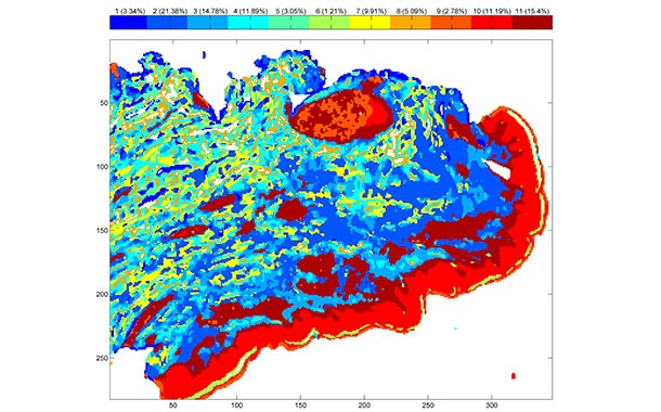
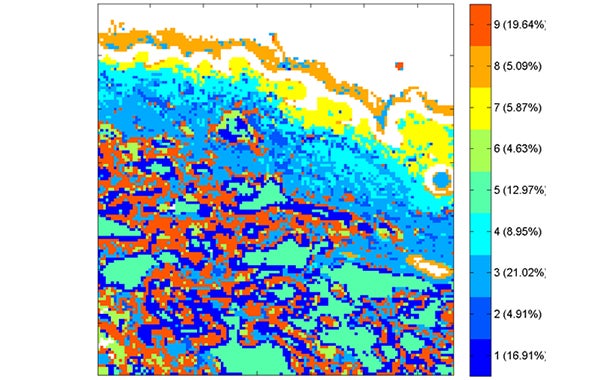
Infrared (IR) spectroscopy
This provides molecular information on skin tissue at a microscopic level. We do this without modifying the sample through staining.
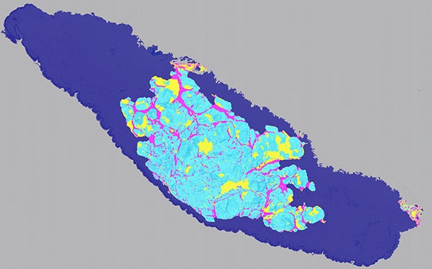
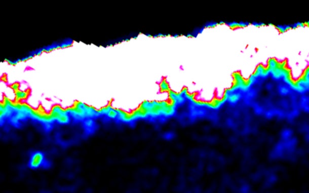
Multi-photon imaging
To investigate skin aging and measure skin collagen, we can generate non-invasive optical biopsies of the skin very quickly, thanks to the development of multi-photon microscopy. We can even picture and completely quantify collagen or other skin constituents.
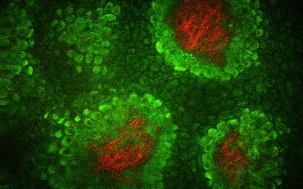
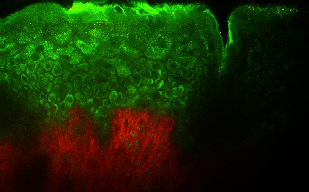
Multi-photons second harmonic generation, Collagen (red), skin dermal papilla fluorescence (green)
MALDI imaging
MALDI (matrix-assisted laser desorption ionisation) is an ionisation technique used in mass spectroscopy to visualise and quantify skin penetration pathways and select efficient formulations. This ground-breaking technology provides a better understanding of compound skin distribution.
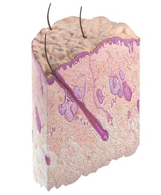
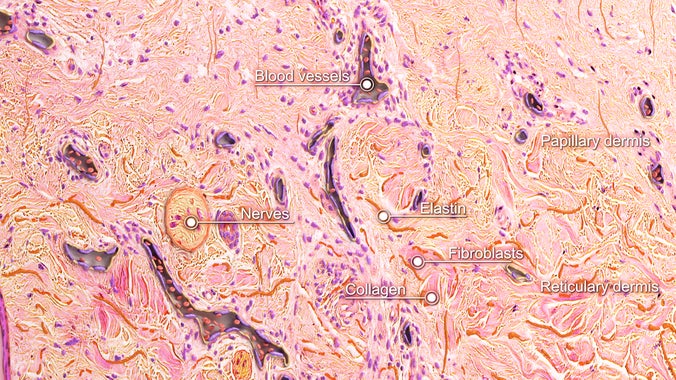
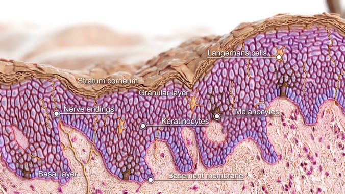
Virtual skin biopsy
The objective of this 3D ‘skin cube‘ is to discover the microscopic structure of normal skin as seen under the microscope, when observing a stained-cut section from a skin biopsy. Skin biopsy is a safe, easy, minimally invasive outpatient procedure of diagnostic and academic relevance. A small amount of skin tissue is removed with a punch, for microscopic examination by a histopathologist. This procedure is routinely used for the diagnosis of skin disease and cancer.